Explaining imaging modalities
Ultrasound

Ultrasound passes sound waves into the body to create pictures from their reflections. This uses no radiation and is therefore safe and provides real-time images but has limitations on the quality of the images obtained.
MRI

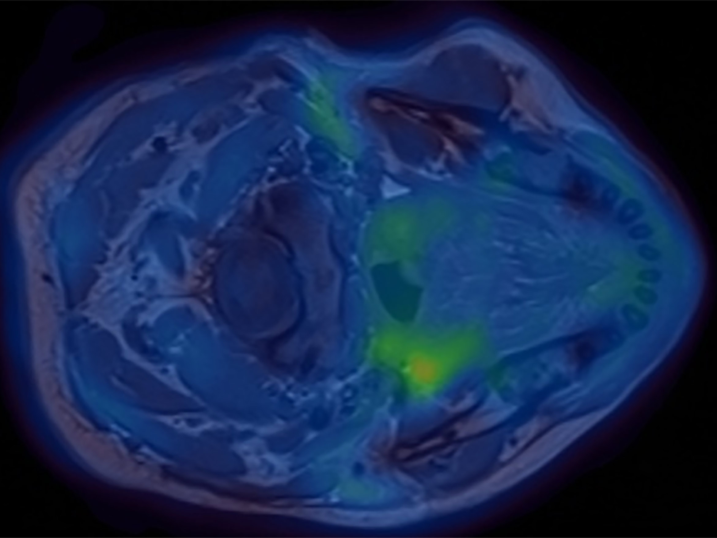
Magnetic resonance imaging (MRI) uses a combination of a strong magnet and radiowaves to produce detailed pictures of the inside of your body. It does not involve the use of either X-rays or radiation. It is particularly good at identifying problems in the spine, the brain, the heart and in the joints.
PET

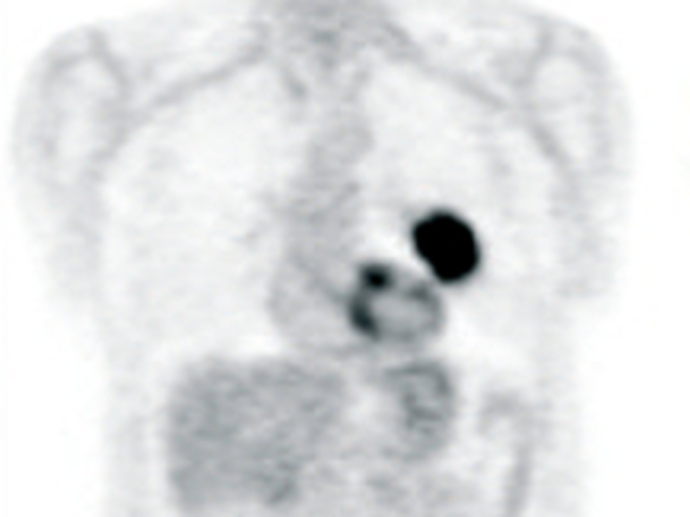
PET (Positron Emission Tomography) requires the injection of a small amount of radioactive tracer, most commonly a sugar, which travels around the body and is taken up by parts of the body that use a lot of sugar. Using the PET scanner we can see the increased use of sugar often associated with disease. We can then localise the exact part of the body using the MRI scan. A PET scan is a very sensitive and accurate method of detecting changes in metabolism associated with diseases, often before abnormalities show up on ordinary scans. PET scans are less effective at showing the exact anatomy of where the abnormality is, this is why there is benefit from combining a PET scan and an MRI scan.
CT

Computerised Tomography (CT) uses X-rays and a computer to create detailed images of the inside of the body. CT scans can produce detailed images of many structures inside the body, including the internal organs, blood vessels and bones.
