Case Studies
TACTIC CRT: study: A prospective randomised multi-centre Trial comparing cardiac MRI guided CRT versus Conventional CRT implantation in patients with Ischaemic Cardiomyopathy.
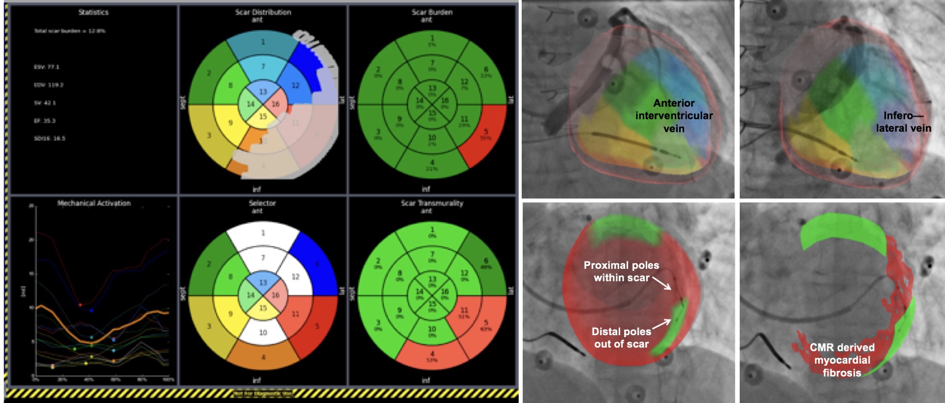
Cardiac resynchronisation therapy (CRT) is an established treatment for improving symptoms and prognosis for patients with heart failure. However, a significant proportion of patients do not respond to due to pacing in scarred tissue and an inability to identify and effectively stimulate those regions of the heart. Image registration/fusion technology alongside computer software to identify the optimal lead location has been co-developed.

The pre-processing workstation of the patient CMR data evaluating scar transmurality and desynchrony is below

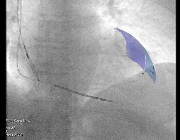
How the final chose segment appears on the real-time fluroscopy
Medical Open Network for AI (MONAI):
Jointly with Nvidia we have released MONAI, an open-source community-supported, deep learning framework for healthcare imaging. MONAI is the only professional-grade platform for medical AI with the quality and features necessary to make better, safer and more robust AI models, which will result in improvements in patient care via improved diagnosis, prognosis, triaging and prioritisation.
MONAI has been downloaded over 50,000 times and it has more than +1.8k starts on GitHub, making it the most popular and used AI software platform for healthcare.
The MONAI consortium is now composed of more than 10 world-leading institutions including Stanford University, UCL, the Chinese Academy of Sciences, the Technical University of Munich and Vanderbilt University.
A New Software Solution for Detecting Heart disease at birth:
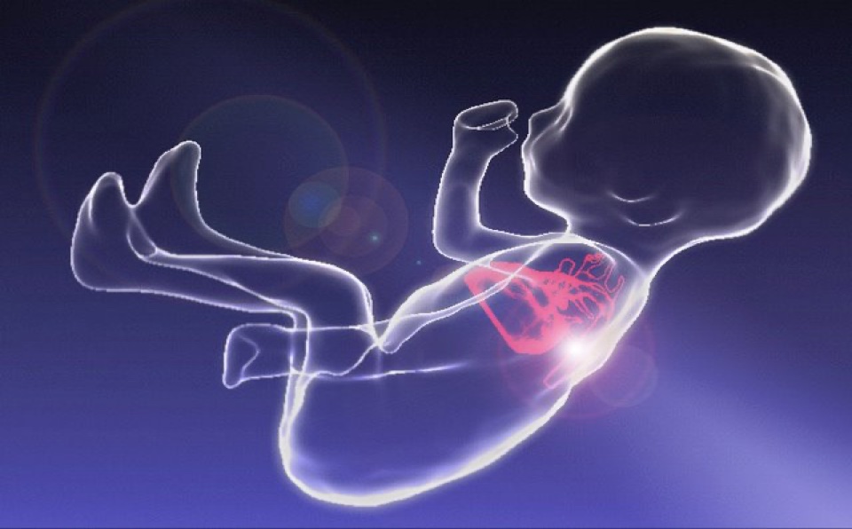
Congenital heart disease is one of the most common types of birth defect, affecting up to eight in every 1,000 babies born in the UK. When congenital heart disease is suspected in a baby before birth, clinicians using ultrasound are not always able to diagnose all details of the condition due to the technical challenges involved with imaging the moving fetus. This is important when abnormalities involve the blood vessels around the heart. 3D imaging of perinatal hearts was developed using MRI and open-source software for 3D motion correction of images [Lloyd et al. Lancet 2019]. 85 patients were enrolled in a prospective study and results showed good alignment with 2-D echocardiography. We now run a clinical service that deploys these methods with approximately three examinations per week.

Galliprost - Democratisation of PET imaging technology for prostate cancer patients through smart radiochemistry:
Prostate cancer kills over 10,000 men annually in the United Kingdom alone. PET imaging is central to managing prostate cancer. Utilising underpinning research from Centre investigators who had developed tris-hydroxypyridinone (THP), a patented chelator for gallium-68, we have helped synthesise a PET tracer using a simple and quick single-vial kit, named Galliprost [Young et al. J Nuc Med 2017]. Galliprost addresses the unmet need for a generic radiolabelling technology that is quick, simple, and cost-effective to use, with minimal infrastructure or expertise. The Centre supported the clinical evaluation of the Galliprost tracer in over 1,000 patients, showing that it meets both its key aims of impacting patient management and being very easily synthesised in the hospital setting [Kulkarni et al J Nuc Med Mol Imag 2020]. This kit has improved treatment decisions in over 1,200 patients globally to date, allowing more appropriate and effective treatment and provided cost savings of £600- £1,500 per patient.

3D coronary MR angiography for non-invasive and radiation-free assessment of coronary artery disease:
Coronary artery disease (CAD) is the leading cause of death world-wide. X-ray coronary angiography remains the clinical gold standard for the assessment of CAD but is invasive and exposes patients to iodinated contrast and ionising radiation. It is limited by complications including death, stroke, myocardial and vascular injury, pain and bleeding. Computed Tomography Angiography (CTA) has emerged as the non-invasive alternative for the anatomical assessment of CAD, but coronary CTA has limited use in certain patient groups. To address the limitations of X-ray and CT coronary angiography a group of researchers embedded within the Centre engineered a novel non-invasive, ionising radiation- and contrast-free coronary magnetic resonance angiography (CMRA) technology for diagnosing CAD [Bustin et al. J Cardio Mag Res 2020]. In its initial testing phase this technology has been successfully used to diagnose CAD in over 200 patients at St Thomas’ Hospital. In 2018, Siemens incorporated this technology in their pre-product package across 10 medical centres worldwide with over 500 patients diagnosed.

89Zr-labelled cell therapy:
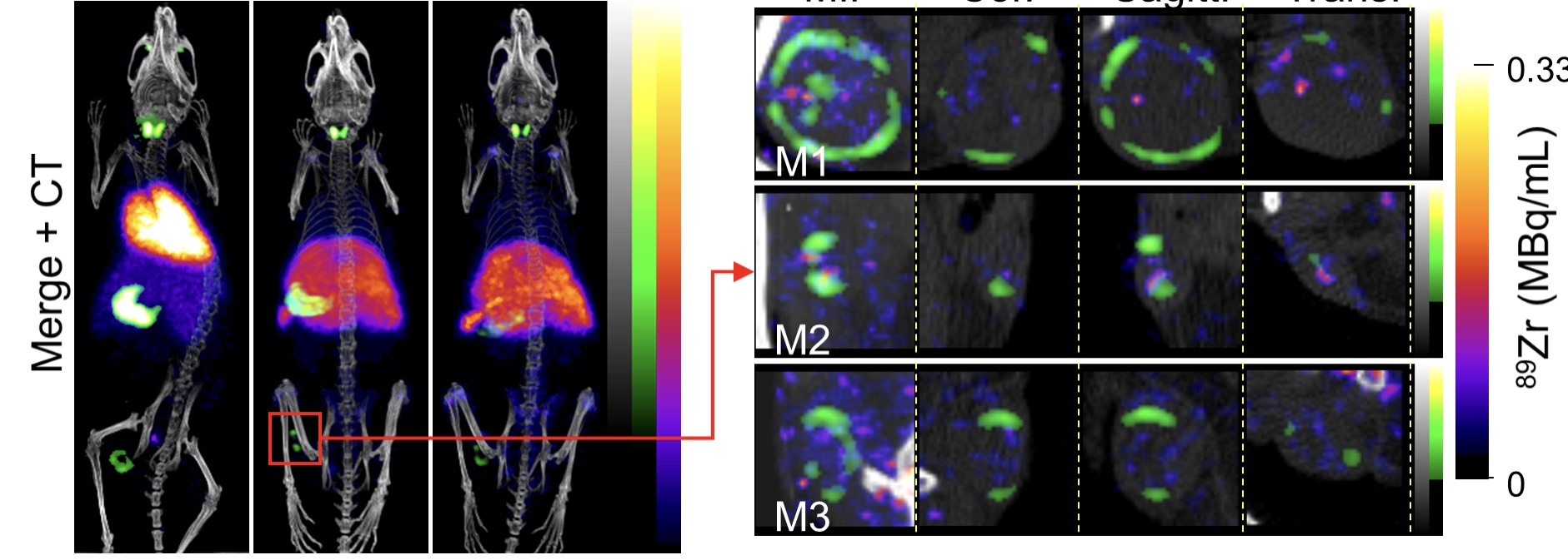
A new and simple kit-based method for preparing 89Zr-oxine for cell labelling has been developed and a patent filed. Funds have been raised, including a contribution from the Centre, to produce the kits commercially to GMP standards. A clinical trial of mesenchymal stem cells (non-labelled) has started as a dose escalation study and the radiolabelled cell element is due to be incorporated later in 2021 and other potential clinical trials are emerging both at KCL/GSST and KCH. A grant has been secured from Research England to work across London sites (QMUL, UCL, KCL) to implement this clinically.

Mouse image of gamma delta T cells – Red = location of cells, green = location of tumour
