Prof Christos Bergeles discusses surgical robotics for eye surgery with Prof Brian Cox as part of a video series on new and emerging STEM technologies for the Royal Society, London.
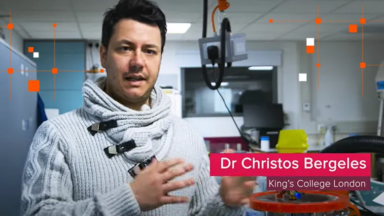
Prof Christos Bergeles discusses surgical robotics for eye surgery with Prof Brian Cox as part of a video series on new and emerging STEM technologies for the Royal Society, London.
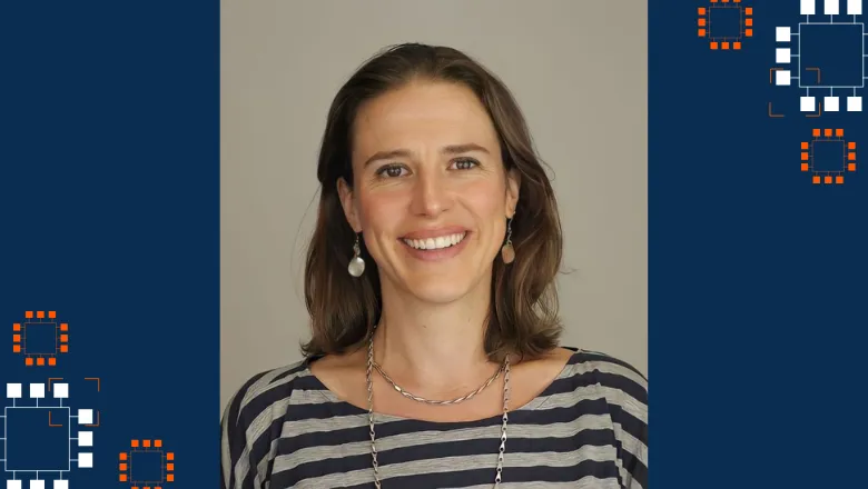
Prof Anne Vanhoestenberghe talks to us about what led her to become Director of MAISI and breaking down barriers for student success.
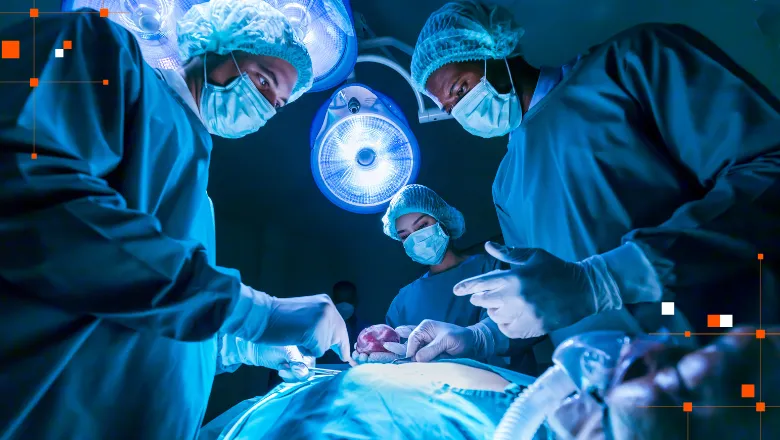
Congratulations to Dr Nicholas Raison, Prof. Prokar Dasgupta, and Dr Alejandro Granados, who have been awarded a grant worth £25,000 by the Royal College of Surgeons of England for their work developing new AI models for robotic surgical skills training.

Jackie is the Design Quality and Regulatory Lead of the Medical Engineering Quality Management System at LIHE. We spoke to her about how her role acts as a bridge between engineering, law and healthcare, and why quality and regulation management is essential to engineering better health.
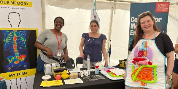
Another year, another Lambeth Country Show with music, vegetable competitions, petting zoos and… BMEIS returns to the Discovery Zone – Science Big Top even BIGGER and BETTER with your help!! We need more demos and more staff to talk to the people.